Plenary session highlights innovative methods for profiling tumor microenvironments in their native tissue context
Monday’s plenary session at the American Association for Cancer Research Annual Meeting 2024 explored innovative approaches to spatially profile not just the morphology and cellular identities but also the multi-omics features, commensal microbial communities, clonal identities, and higher-order organization of the tumor ecosystem.
The session, Profiling Tumor Ecosystems in Native Tissue Context, can be viewed on the virtual meeting platform by registered Annual Meeting participants through July 10, 2024.

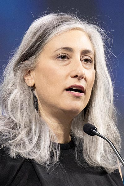
Session Chair Christine A. Iacobuzio-Donahue, MD, PhD, director of the David M. Rubenstein Center for Pancreatic Cancer Research at Memorial Sloan Kettering Cancer Center, emphasized that tumors do not exist in isolation.
“They are not cells in a dish. They are not organoids growing in a 3D dome of Matrigel. They are cellular compositions that are at interaction with many other types of cells,” Iacobuzio-Donahue said. “They may be located in different parts of the bodies and exposed to a huge variety of cellular components that influence how they behave.”
Spatial encoding of immune response

Michael Angelo, MD, PhD, assistant professor of pathology at Stanford University School of Medicine, focused on the development and application of a multiscale, multimodal framework for mapping the spatial biology of the tumor microenvironment (TME) that integrated three techniques—spatially resolved proteomics to map 40–50 proteins; spatially resolved transcriptomics to query expression of hundreds to thousands of targets; and spatial glycomics to map tissue glycans.
He illustrated how this framework could provide insights on the complex tumor ecosystem using three case studies—glioblastoma multiforme (GBM); ductal carcinoma in situ (DCIS) of the breast; and metastatic triple-negative breast cancer (mTNBC).
“We can use these techniques to inform drug regimens in GBM, we can use them to understand from a topological perspective why some people seem to progress to invasive disease from DCIS, and lastly, we think we’re going to be able to use these approaches to predict responses in drug therapy,” Angelo said.
Understanding the impact of intratumoral microbes

Susan Bullman, PhD, assistant professor at Fred Hutchinson Cancer Center, explored how tumor-associated microbial communities interact with tumors and the potential impact of microbial niches, or “microniches,” on tumor behavior. Different bacterial taxa have been shown to be enriched within tumor tissue compared to the microbes found in normal tissue. For instance, in gastrointestinal tract (GI) tumors, Fusobacterium nucleatum is enriched; moreover, intratumoral enrichment of these bacteria is associated with unfavorable outcomes.
Bullman presented findings from analyses of the microbial milieu in GI tumors, including oral cavity squamous cell and colorectal cancer, using spatial transcriptomics and unbiased molecular mapping of microniches. Bullman also elucidated on the development and validation of a host-microbial, single-cell RNA sequencing platform.
She said the data demonstrated that microbes are not distributed evenly within a patient’s tumor. Instead, their distribution is incredibly heterogenous but highly organized to form microniches that impact and interact with both cancer epithelial cells and immune cells that may promote cancer progression.
“Moving forward, we really need to think about how we can harness this new information on the tumor microenvironment and harness these new technologies to think about what these microbes are actually doing within patient specimens and how they are contributing to cancer progression and impacting patient treatment response,” Bullman said.
Deconvoluting the cancer ecosystem into its spatial components

Joakim Lundeberg, PhD, professor at KTH Royal Institute of Technology and Science for Life Laboratory, both in Sweden, presented on the development and application of a spatial transcriptomics approach, based on three core pillars—the capture of whole transcriptome data in a given tumor section, including spatial information; data-driven analysis to enable spatial clustering based on similarities in gene expression profiles; and spatial visualization of gene expression data across the tissue section.
Lundeberg stated that their aim is to maximize the information that can be gathered from each tissue section. Spatial transcriptomics, along with mRNA and DNA sequencing and metabolomic analyses, provides granular information on spatial differences within tumor tissue sections. Such spatially resolved analyses can help identify minor subsets or small populations of cells within tumors with distinct characteristics, that are not readily identifiable or captured using other methods, Lundeberg noted.
Dissecting plasticity in tumor cell states in primary melanoma

Peter K. Sorger, PhD, Otto Krayer Professor of Systems Pharmacology, Department of Systems Biology, Harvard Medical School, gave an example of novel high-resolution 3D tissue profiling approaches, applied to melanoma. He spoke about the disadvantages of profiling approaches that use thin sections compared to the “true 3D” structure that can be illuminated using thicker sections.
Sorger focused on the application of thick specimen optical sectioning and cyclic light sheet fluorescence microscopy. In precancerous melanoma, their 3D imaging studies provided a picture of the association between inflammatory signatures and initiation of T-cell migration. In metastatic melanoma, Sorger described the high degree of heterogeneity across all levels of spatial analyses—metastatic lesions were spatially heterogeneous in terms of their morphology, cell type distribution, gene expression programs, and clonal distribution. Moreover, the organization of cells within metastatic tumors centered around macroscopic features, such as vasculature or peri-necrotic regions, rather than around their granular characteristics. Sorger said that these innovative spatial profiling methods allow better correlation of genotype to phenotype within the TME.
Register Today for the AACR Annual Meeting 2025
Don’t miss the most important cancer meeting of the year, April 25-30 in Chicago, Illinois. In-person and virtual registration packages include full access to live sessions, Q&A, networking, CME/MOC credits, and more.

