Plenary speakers explore the latest advances in the early detection and interception of cancer
During the plenary session Early Detection and Interception of Cancer, five researchers described multiple avenues of research and discovery that demonstrate how cancer can be detected and assessed early to improve patient outcomes.
The session, which was originally presented Monday, April 17, can be viewed on the virtual meeting platform by registered Annual Meeting participants through July 19, 2023.

Phil H. Jones, PhD, Professor of Cancer Development at the University of Cambridge and Senior Group Leader at the Wellcome Sanger Institute, United Kingdom, described how the mutational landscape of the esophagus may relate to cancer risk and explored whether it’s possible to intervene to halt the expansion of oncogenic mutant clones.
“We are progressively colonized by mutant clones as we age, and the aging esophagus becomes a Darwinian battleground in which mutant clones fight for space, and only the fittest survive,” Jones said. “These mutations are almost all due to cell-intrinsic process that correlate with age, so are inevitable and inexorable.”
The ability to outcompete other mutants in normal tissue, however, does not correlate with carcinogenic potential. For example, NOTCH1 mutant clones colonize most of the esophagus by middle age, but NOTCH1 mutations are comparatively rare in squamous cancers and seem to be anti-oncogenic, removing nascent tumors and inhibiting the growth of larger tumors, Jones explained.
In contrast, studies have shown that mutant TP53 is less effective than NOTCH1 at driving clonal expansion but is found in almost all human squamous esophageal cancers and contributes to carcinogenesis in mice.
“So, if NOTCH1 is a desirable mutant, TP53 is a bad one,” Jones said. “If we can understand the phenotype of expanding oncogenic mutant clones, it might be possible to halt their expansion by interventions — either by altering the environment or with pharmacological agents — by leveling out phenotypic differences, or even by making oncogenic mutant cells less fit than their neighbors so they are depleted from the tissues, thus reducing cancer risk.”

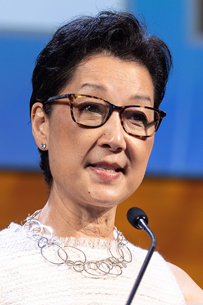
E. Shelley Hwang, MD, MPH, Professor of Surgery and Vice Chair of Research in the Department of Surgery at Duke University School of Medicine and the Duke Cancer Institute, explained how genomic profiling of accurately annotated longitudinal case control datasets have provided insights into breast lesions known as ductal carcinoma in situ (DCIS), which are thought to be precursors to invasion and metastases.
Both transcriptomics and spatially resolved proteomic characterization of DCIS have uncovered novel biology of the epithelial cells that make up DCIS, as well as the heterogeneous precancer ecosystems that can be either permissive or inhibitory for invasion. Recent work has also uncovered a clonal evolution pathway characterized by multiclonal invasion, which has helped researchers build risk-stratification tools to refine treatment recommendations.
“For asymptomatic tumors at low risk of cancer progression, there may be little to no benefit to treatment, whereas for more high-risk lesions, progression to invasion and metastasis may be rapid and deadly,” she said.
Given the time between the development of precancer and progression to invasive disease, Hwang said there may be a case for tailoring intervention of screen-detected cancer by age and the presence of competing comorbidities.
“The decision of whether and when to intervene for screen-detected cancers will require a multidisciplinary effort, including expertise in cancer biology, mathematical modeling, psychology, health policy, decision science, and communication, as well as thoughtful engagement from patients and health care providers to identify the optimal time to intervene along the cancer progression continuum,” Hwang said.

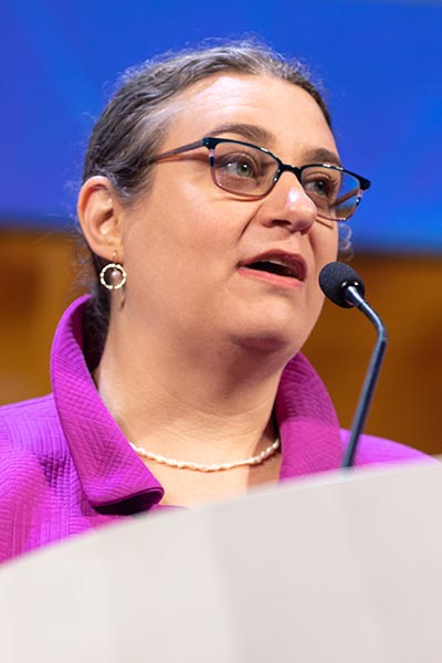
Elana Judith Fertig, PhD, Professor of Oncology and Division Director of Oncology Quantitative Sciences at Johns Hopkins University and the Sidney Kimmel Comprehensive Cancer Center, discussed a spatial multi-omics approach to study pancreatic cancer development — namely the interactions between neoplastic cells and the microenvironment during pancreatic carcinogenesis.
New single-cell and spatial molecular profiling technologies have enabled unprecedented characterization of the cellular and molecular composition of the microenvironment in advanced pancreatic cancer, which includes a dense composition of cells that are associated with immunosuppression. The convergence of these technologies with machine learning and mathematical modeling, Fertig said, provide the potential to identify candidate therapeutics to intercept immunosuppression in pancreatic cancer.
“Combining genomics with mathematical modeling provides a forecast system that can yield computational predictions to anticipate when and how the cancer is progressing for therapeutic selection,” Fertig said. “This mathematical forecast system will empower a new predictive oncology paradigm, which selects therapeutics to intercept the pathways that would otherwise cause future cancer progression.”

Phillip G. Febbo, MD, Chief Medical Officer at Illumina Inc., addressed the complexity of early cancer detection at the societal level and the potential to improve cancer outcomes through equitable access to testing, including emerging multi-cancer early detection tests (MCEDs).
“Most lethal cancers have no screening test. It’s estimated that as much as 70 percent of cancer deaths are due to cancers without available screening,” he said. “MCED testing has the potential to transform cancer detection by identifying cancers at earlier stages when they may be more treatable.”
MCED screening could reduce cancer mortality by 20 percent, said Febbo, citing findings from the PATHFINDER study in which MCED testing more than doubled the number of cancers detected with recommended screening.
“Equity of access and adoption, however, are critical. By some estimates, disparities in development and/or adoption of MCED screening could reduce the benefit by 50 percent,” he said.

Karriem S. Watson, DHSc, MS, MPH, Chief Engagement Officer of the All of Us Research Program at the National Institutes of Health, highlighted the importance of community engagement in early cancer detection strategies as a key to improving patient outcomes.
Studies have demonstrated that community engagement can help mitigate cancer health disparities by increasing communities’ knowledge of the importance of healthy behaviors, building trust in research, and improving study design and ethics, he said. Increasing access and awareness of medical studies with an emphasis on populations that have been historically underrepresented in biomedical research (UBR) has also been shown to expand the reach of innovative and novel breakthrough therapies for early detection and improve cancer outcomes.
“Engaging UBR communities in the early phases and throughout the duration of research studies is important to identify the right problems, formulate the appropriate questions, and build mutual trust and transparency,” Watson said. “Researchers also play an essential role in community engagement, as studies have shown that being engaged by a research team that reflects the diversity of the population being studied leads to greater participation in research, better retention, and adherence to recommendations.”
Register Today for the AACR Annual Meeting 2025
Don’t miss the most important cancer meeting of the year, April 25-30 in Chicago, Illinois. In-person and virtual registration packages include full access to live sessions, Q&A, networking, CME/MOC credits, and more.

